Schwannoma

Thank you for visiting my blog. My name is Dr. Ramin Rak and I am a surgeon with Neurological Surgery, P.C.

I specialize in treating complex spinal and brain complications at the eleven different medical facilities affiliated with Neurological Surgery, P.C. and I am a member of the organization’s Neurosurgeon Brain Tumor Team.

The Brain Tumor team includes an Endovascular Neuroradiologist, two Neuro-oncologists, a Neuropsychologist, myself, and seven other neurosurgeons:

  • Dr. Michael H. Brisman, M.D., F.A.C.S.
  • Dr. Jeffrey A. Brown, M.D., F.A.C.S.
  • Dr. Lee Eric Tessler, M.D., F.A.A.N.S.
  • Dr. Alan Mechanic, M.D., F.A.C.S.
  • Dr. Robert N. Holtzman, M.D.
  • Dr. Vladimir Dadashev, M.D.
  • Dr. Gerald M. Zupruk, M.D., F.A.A.N.S.

The rest of the Brain Tumor Team and I work to treat brain tumors and complications caused by tumors, including gioblastoma, medulloblastoma, acoustic neuroma, and schwannoma.

Schwannoma is a homogenous tumor that is made up entirely of Schwann cells, which conduct nervous impulses along axons, provide trophic support for neurons, and support nerve development and regeneration.

The tumor cells are always on the exterior of the nerve though the tumor itself can cause nerve damage by pushing the nerve aside or into bone. Schwannomas become malignant in less than 1% of cases and are slow growing, but should still be treated if weakness numbness, pain or other symptoms are seen.

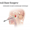
The Brain Tumor Team at NSPC uses surgery or stereotactic radiosurgery to treat schwannomas with radiosurgery being an option when the tumor is located in the head or spine.

The most common treatment is stereotactic radiosurgery, or the use of a machine called the Gamma Knife to focus high-powered x-rays at the spot of the tumor. Between 80 and 90% of the patients who opt instead to have surgery report that pain, weakness, and numbness disappears following the procedure.

Learn more about how the Brain Tumor Team treats schwannomas here.

Thank you for reading,

Dr. Ramin Rak


Ramin Rak Schwannoma

This is an example of a schwannoma. It typically has dense areas called Antoni A (black arrow) and looser areas called Antoni B (blue arrows). The cells are elongated (spindle shaped) and the nuclei have a tendency to line up as you see here in the Antoni A area. (via ucsf.edu)

NSPC Brain Tumor Center

NSPC Brain Tumor Center with Ramin Rak

Hi all, Ramin Rak here with another blog post about my work as a neurosurgeon.

I am affiliated with Neurological Surgery, P.C., New York’s private largest neurosurgical group. One of the resources that Neurological Surgery, P.C. uses to provide better treatment is the Long Island Brain Tumor Center (their official website is located here).

I am one of the neurosurgeons who is part of the Neurosurgeon Brain Tumor Team, along with:

  • Dr. Michael H. Brisman, M.D., F.A.C.S.
  • Dr. Jeffrey A. Brown, M.D., F.A.C.S.
  • Dr. Lee Eric Tessler, M.D., F.A.A.N.S.
  • Dr. Alan Mechanic, M.D., F.A.C.S.
  • Dr. Robert N. Holtzman, M.D.
  • Dr. Vladimir Dadashev, M.D.
  • Dr. Gerald M. Zupruk, M.D., F.A.A.N.S.

The Brain Tumor Team also includes an Endovascular Neuroradiologist (Dr. John Pile-Spellman, M.D.), a Neuropsychologist (Dr. Gad E. Klein, Ph. D.), and two neuro-oncologists (Dr. Paul Duic, M.D. and Dr. Jai Grewal, M.D.).

The Long Island Brain Tumor Center focuses on treating conditions like acoustic neuroma, schwannoma, ependymoma, astrocytoma, and other ailments related to brain tumors.

A brain tumor occurs as a result of uncontrolled cell division in the brain itself, glial cells, lymphatic tissue, the cranial nerves, brain envelopes, the skull, pituitary and pineal gland, or can even be spread from cancer located in other areas. Symptoms of a brain tumor include phantom odors and tastes, while onset symptoms like an epileptic seizure in a patient with no history of epilepsy or sudden intracranial hypertension have also been observed.

The team of neurosurgeons within the Long Island Brain Tumor Center diagnoses tumors using computed tomography (CT) and magnetic resonance imaging (MRI) mapping techniques.

Tumors must be investigated before a diagnosis and treatment options can be determined, but the good news is that not all brain tumors are cancerous.

Take a look at my other blog posts to find out how I use techniques like Gamma Knife, and CyberKnife procedures to treat brain tumors without surgery.

Thanks for reading,

Ramin Rak

Spinal Fusion

Good afternoon, and thank you for visiting my blog.

My name is Dr. Ramin Rak and I am a neurosurgeon with Neurological Surgery, P.C. Though I primarily specialize in techniques that treat brain injuries and tumors (like the Cyber Knife and GammaKnife procedures that I spoke of in earlier blog posts), I also complete complex procedures intended to alleviate spinal conditions. Last week I detailed my use of the X-Stop Procedure for individuals suffering from back pain and this week I will talk about my technique for treating a condition called spondylolisthesis.

Spondylolisthesis occurs when a damaged or fractured joint allows a vertebra to slip forward and pinch a nerve joint in the spine.

This pain will travel all the way down to the patient’s legs via the sciatic nerve. Surgeons perform a spinal fusion to treat this condition and alleviate patient pain. At Neurological Surgery, P.C. I am a member of the Spinal Fusion Team along with:

  • William J. Sonstein, M.D. F.A.C.S.
  • Benjamin R. Cohen, M.D. F.A.C.S.
  • Artem Y. Vaynman, M.D. F.A.A.N.S.
  • Donald S. Krieff, D.O. F.A.C.O.S.

My team and I perform the spinal fusion procedure to treat intractable back pain by fusing two or three lumbar spine segments.

We start by removing the lamina, which is the portion of the spine that covers the problematic vertebra. Next, the Spinal Fusion team removes any piece of bone that is pinching the vertebra, relieving any pressure and pain. A bone graft is then performed, which fuses together the two spine segments around the vertebra. Finally, screws and rods are used to hold the spine in place while the grafts heal. Once the grafts heal, they will fuse into place and keep the discs from slipping and causing future complications.

Learn more about the spinal fusion team and the PLIF (posterior lumbar interbody fusion) or spinal fusion procedure by clicking here to view a video demonstration on NSPC’s website. Stay up to date on my other neurosurgical techniques when you follow me on Twitter.

Thanks for reading,

Dr. Ramin Rak


Ramin Rak Spinal Fusion

Screws and rods are used to hold the bone grafts in place so that the spinal fusion results are permanent.

The X-Stop Procedure

Hello all, Dr. Ramin Rak here with another blog post.

Many of my posts have focused on complex neurosurgical techniques that I have used and still use, but I also specialize in completing complex spinal surgeries for individuals suffering from back pain. One of my specialties is the X-Stop Procedure, and I am one of only seven surgeons at Neurological Surgery, P.C. who complete this procedure. The others are Dr. Stephen D. Burnstein, Dr. William Sonstein, Dr. Benjamin Cohen, Dr. Artem Vaynman, Dr. Alan Mechanic, and Dr. Donald Krieff.

The X-Stop Procedure involves the insertion of a titanium implant called the X-Stop Spacer.

The spacer is placed in order to treat back pain and leg pain caused by lumbar spinal stenosis (LSS). LSS takes place when space between the vertebrae is reduced, as this lack of space causes bone or tissue to come in contact with the spinal nerve. Individuals who are suffering from LSS find relief when seated or bending over because these positions open up space between the vertebrae. The X-Stop Procedure draws inspiration from this concept, using the X-Stop Spacer to lift the vertebrae off of the pinched nerve and provide long-term relief.

X-Stop Spacer

A diagram of the X-Stop Spacer following placement.

I complete the minimally invasive X-Stop Procedure by surgically inserting the X-Stop Spacer between two bones in the back of the spine; usually L3/4 or L4/5.

The surgical site is located between the posterior spinous processes, which is an area you can feel for yourself by running a finger along your spine. The X-Stop Spacer stays in place by working with one’s natural spinal anatomy, which means that the procedure is completed without using screws or needing to be attached to bones or ligaments.

The X-Stop Procedure takes between 50 and 90 minutes, and patients are able to walk around shortly after the anesthesia wears off. Patients see results immediately after the procedure, as they will no longer experience back pain when standing or walking around.

Learn more about the X-Stop Procedure by visiting their website, or leave a comment below if you still have questions.

Thank you for reading,

Dr. Ramin Rak

My Second Awake Craniotomy

Good afternoon and welcome back to my blog.

My name is Ramin Rak, I am a neurosurgeon with Neurological Surgery, P.C. in New York and one of my specialties is performing awake craniotomies.

In July of 2010, I received media attention after completing the first awake craniotomy that had ever been performed at the North Shore-LIJ Huntington Hospital.

The procedure was notable for me because it was only my second awake craniotomy.

The need for the procedure was uncovered after 23-year-old Boris Arrazia experienced a seizure while driving his car. Arrazia’s passenger was able to navigate the car to the side of the road and put it in park, and a passerby performed CPR and then called for help.

Arrazia was brought to Huntington Hospital where doctors uncovered the cause of the stroke: a tumor located in the temporal lobe of his brain.

Due to the tumor’s unusual location, the tumor did not show up when using advanced imaging techniques. Because the tumor was right on his speech area, there was no way to map and understand how we are going to control the reception of the brain without the patient being awake. By performing an awake craniotomy, my team was able to make sure we did not damage areas of the brain dealing with speech during surgery by asking Arrazia to repeat phrases or describe pictures. Arrazia recounts:

“I was nervous when Ramin Rak first told me the diagnosis, but happy to hear that the tumor was operable,” Arriaza said. “Ramin Rak told me that I would be conscious for the surgery, and that it might be uncomfortable and possibly a little painful. During the surgery, they showed me pictures and asked me to name the objects [like] a horse [or] a table.”

Though there are a lot of risks associated with awake craniotomies, including serious bleeding, there were no complications seen while removing Arrazia’s tumor. Four days later, he walked out of the hospital and reunited with his family.

Learn more about this particular awake craniotomy by taking a look at the media coverage this procedure received:

http://www.northshorelij.com/NSLIJ/Islip+Man+Undergoes+Brain+Surgery+While+Awake  

http://huntington.patch.com/articles/huntington-hospital-doc-performs-awake-craniotomy


If you are interested in learning more about how I perform awake craniotomies, visit my Quora profile.

Thank you for reading,

Ramin Rak

Ramin Rak awake craniotomy

Boris Arrazia thanks Ramin Rak following completion of the awake craniotomy.

Leslie Munzer Neurological Institute

Hi all, Dr. Ramin Rak here with another blog post about neurosurgery.

Some of my past blog posts have focused on complex neurosurgical techniques that I use, but I do not spend all of my time in the operating room. Part of my success can be attributed to the education along the way, from my eight years of undergraduate and medical schooling at the Free University of Brussels to the countless residencies I completed in hospitals throughout the United States. For this reason, I make it a priority to give back to the medical community by educating them on the techniques that I use. One way that I do so is by keeping up this blog, but I also present lectures at medical seminars across the country.

Back in 2009 I was asked by the Leslie Munzer Neurological Institute (LMNI) to speak at a comprehensive educational seminar on Brain Tumor and Stroke Awareness.

This two hour seminar took place at the Jefferson’s Ferry Lifetime Retirement Community’s Community Center in New York, and lecture topics included, “Stroke- Treatment, Recovery, and Prevention,” “Current Treatments of Brain Tumors,” “A Stroke Overview,” and “New Chemotherapy Options for the Treatment of Brain Tumors.” My lecture was titled “Advancements in Brain Tumor Surgeries” and I discussed advanced microneurosurgical techniques like awake craniotomies, the Gamma Knife procedure, and the CyberKnife system.

The Leslie Munzer Neurological Institute was formed in 2006 by Neurological Surgery, P.C. and at the time was called The Long Island Neurological Institute, Inc.

This organization coordinated funds for research related to the brain and spinal cord while supplying information to patients and the medical community about neurological ailments.

The organization changed its name in 2008 in memory of Leslie Munzer, who passed away in 2004 from a ruptured arteriovenous malformation (AVM).

Shortly after her passing, the Munzer family had formed a charity in her name to raise funds for AVM research and awareness, and in 2008 donated the collected funds to The Long Island Neurological Institute, Inc.

You can learn more about my efforts to educate the neurosurgical community with The Leslie Munzer Neurological Institute and with other organizations by viewing my other blog posts.

Thank you for reading,

Dr. Ramin Rak

Ramin Rak at May 2009 Seminar

Dr. Ramin Rak (second from the right) and the other speakers asked to present a lecture for the May 2009 Leslie Munzer Neurological Institute (LMNI) seminar.

Gamma Knife Procedure

Hello readers, Ramin Rak here with yet another blog post about a complex neurosurgical method that I have used to treat patients.

Gamma Knife is a procedure, like the CyberKnife System, that allows doctors like me to treat tumors and ailments without having to resort to surgery. Gamma Knife surgery was approved for use by the US Food and Drug Administration (FDA) thirty years ago and I am one of the few New York area neurosurgeons certified in this technique.

When I use Gamma Knife procedures for a patient, I can deliver over 200 beams of radiation right to the tumors and lesions.

Unlike with traditional radiation, patients do not experience the regular side effects because such low doses of radiation are injected. Patients can be given several shots of radiation during one session, and sessions can be repeated until the tumor has stopped growing.Gamma knife procedures can be used to treat:

  • Benign tumors, such as meningiomas, schwannomas, craniopharyngiomas, pineal tumors, acoustic neuromas, and pituitary adenomas.
  • Malignant tumors, such as metastatic tumors, chordomas, ependymomas, medulloblastomas, astrocytomas, and anaplastic astrocytomas.
  • Arteriovenous malformations (ATV)
  • Trigeminal neuralgia
  • Parkinson’s Disease

Generally Gamma Knife use is restricted to those tumors that are less than 4 centimeters in size.

This procedure has a success rate between 90 and 95 percent, as that is how often the procedure stops tumor growth. The majority of the time it also causes tumors to shrink, taking anywhere from one week to a year to stop growth.

When a patient undergoes Gamma Knife treatment, he or she will be fitted with a clear, plastic frame.  My team uses 3D computer imaging to determine where radiation must be aimed to successfully reach the tumor. Patients are then fitted with the Gamma Knife apparatus, which is a metal helmet that emits radiation beams. The apparatus then directs radiation beams to the tumor.

This is how I complete Gamma Knife treatments. If you have any questions, check to see if I have answered them on Quora, or reach out to me directly.

Thanks for reading,

Ramin Rak