Symptoms of a Stroke

F.A.S.T. Stroke Symptoms

Ramin Rak breaks down the F.A.S.T. stroke symptoms.

Ramin Rak practices his advanced knowledge of microneurosurgical techniques at Neurological Surgery P.C. on Long Island.

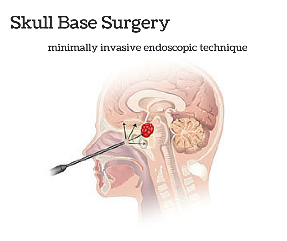
Here, he specializes in awake craniotomies, spinal surgeries, skull-based surgeries, and treatment of tumors.

More information on these complex procedures can be accessed through his Pinterest page.

A stroke is caused by a disturbance in the blood supply and results in the loss of function.

An ischemic stroke is caused by a clot that obstructs blood flow, while a hemorrhagic stroke is a result of a rupturing blood vessel. A transient ischemic attack can also occur, which is essentially a mini stroke caused by a temporary clot. Both types of strokes are extremely serious, as they are the number four cause of death, and the leading cause of adult disability in the United States.

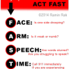
According to strokeassociation.org, F.A.S.T is an easy way to remember the sudden signs of a stroke:

  • Face Drooping- If one side of your face is drooping or your smile is uneven, it could indicate a stroke.
  • Arm Weakness- When one arm is weak, numb, or drifts downward when your arms are raised, seek immediate medical attention.
  • Speech Difficulty- If someone is struggling to form words or unable to repeat a simple sentence, a stroke may be the cause.
  • Time to call 911- If you are experiencing any of these symptoms, call 911 immediately, even if the symptoms go away.

Beyond these core symptoms, a person may also present numbness of the leg, arm or face; trouble seeing; an unexplained sudden, severe headache; dizziness or loss of coordination; and confusion or trouble understanding.

Quick stroke treatment can save lives, so it is crucial to seek immediate medical attention if you have shown any of the above symptoms. After a doctor makes a diagnosis, they will begin a treatment plan specific to the cause of the stroke.

Skull Base Surgery

Ramin Rak is a board-certified neurosurgeon with years of experience specializing in complex tumors, spine treatments, awake craniotomies, and skull base surgeries.

Skull base surgery is a specialized type of surgery performed to treat tumors and other diseases occurring in certain areas of the skull such as behind the eyes or in the nasal cavity. The skull is made up of bones and cartilage, forming the face and the cranium. The bones that form the base of the cranium also form the eye socket, some of the sinuses, the roof of the nasal cavity, as well as the bones surrounding the inner ear. The base of the skull is a very complex area where many vital parts of the body pass through including blood vessels, nerves, and the spinal cord.

Ramin Rak | Skull Base Surgery

Ramin Rak uses the latest in minimally invasive techniques to perform skull base surgery.

Skull Base Surgery requires a multidisciplinary approach often involving ear, nose, and throat surgeons, neurosurgeons, plastic surgeons, radiologists, pathologists, oncologists, and other specialists. There are two main ways to perform skull base surgery. It may be done through a minimally invasive endoscopic procedure. This involves making a small incision through the natural openings of the skull, normally the nose or mouth. A small hole may also be made just above the eyebrow. Through this method, a neurosurgeon is able to remove the growth through a thin lighted tube referred to as an endoscope. An MRI picture of the skull base will usually be taken during this process to ensure that the entire growth has been removed successfully. The second way to perform skull base surgery is often referred to as traditional or open skull base surgery. This procedure requires making an opening in the skull. Parts of bone may be removed in order to reach the growth and have it properly removed.

Listed below are some of the common growths and conditions that may be treated with skull base surgery:

  • Growths caused by infections
  • Pituitary tumors
  • Sinus tumors
  • Glomus tumors
  • Cysts developed from birth
  • Cerebrospinal fluid leak
  • Meningiomas
  • Chordomas
  • Trigeminal neuralgia
  • Acoustic neuroma

Meningitis Symptoms

Thank you for visiting my (Dr. Ramin Rak’s) medical blog!

Today we will be going over the life-threatening condition known as meningitis.

Meningitis SymptomsThe swelling many times identified with Meningitis triggers the early symptoms of fever, headache, and sore neck.

In many cases, the reason for meningitis stems from viral infections.

However, sometimes bacterial and fungal infections can cause meningitis. Depending upon the cause of the inflammation and the associated infection, meningitis conditions can be as simple as waiting a few days, or in some severe cases, can require immediate emergency care. People experiencing symptoms of meningitis are encouraged to see a doctor as soon as possible to safeguard against life threatening conditions.

Meningitis is fought off by antibacterial medication.

Early treatment of bacterial meningitis can prevent serious complications. I urge anyone experiencing the following symptoms to seek medical care immediately. Visit http://about.me/DrRaminRak for more info about my medical background.

  • Sudden high fever
  • Severe headache that isn’t easily confused with other types of headache
  • Stiff neck
  • Vomiting or nausea with headache
  • Confusion or difficulty concentrating
  • Seizures
  • Sleepiness or difficulty waking up
  • Sensitivity to light
  • Lack of interest in drinking and eating
  • Skin rash in some cases, such as in meningococcal meningitis

Symptoms in newborn babies are presented differently.

Please be on the lookout for these symptoms of early meningitis. If your newborn baby diagnosed with meningitis, I recommend parents seek professional counseling simultaneously with the newborn’s treatment and recovery period. The inability to comfort the newborn mentally can be burdening to any parent.

Look for these early signs:

  • High fever
  • Constant crying
  • Excessive sleepiness or irritability
  • Inactivity or sluggishness
  • Poor feeding
  • A bulge in the soft spot on top of a baby’s head (fontanel)
  • Stiffness in a baby’s body and neck

Always seek medical care first before self-prescribing medications.

Any of the listed symptoms could be signs of many of other medical conditions.  A misdiagnosis could lead to life-threatening implications. Visit my website or Tumblr for more information.


Neurological Complications of Lyme Disease

Neurological Complications of Lyme Disease

Ramin Rak explains the Neurological complications of Lyme Disease.

As a highly skilled board certified neurosurgeon, Ramin Rak has treated a number of conditions involving both the spine and brain.

At Neurological Surgery, P.C., located on Long Island, he has come across a number of cases of Lyme disease. With the condition on the rise, it is important to know how lyme disease is contracted and the damaging effects it can have. More tips like these can be found on Ramin Rak’s twitter feed.

According to an article from Stony Brook Medicine, Lyme disease is an infection caused by the bacterium, spirochete.

It is transmitted by deer ticks, which are found throughout North America, Europe, and Asia. A tick bite, though painless, can cause arthritic complications, as well as neurological and cardiac problems. The longer the tick is attached, the greater risk of Lyme disease transmission.

The symptoms and severity of Lyme disease can vary from person to person.

The most common symptom is a rash, which eventually grows into a bulls-eye shaped ring. Flu like symptoms are also possible, including headache, aching muscles, fatigue, chills, and fever. If treated properly, the complications may end there. Unfortunately, if the tick goes unnoticed, a persons first symptoms may be arthritic, neurological, or cardiac.

Arthritic complications due to Lyme disease include general achiness, pain, and swelling in the joints.

Neurological problems consist of meningitis, memory loss, fatigue, difficulty concentrating, bell’s palsy, and encephalitis. Most patients with flu like symptoms can be completely cured with antibiotics but those with arthritic or neurological symptoms require a more vigorous treatment.

Protect yourself from ticks by:

  • Applying tick repellants
  • Shampoo and shower after being in the woods
  • Keep long hair tucked away
  • Wear long sleeves and long pants
  • Check yourself occasionally
  • More tips on how to defend yourself against ticks can be found here.

Learn more about Ramin Rak and the services he provides by visiting wordpress.com.

Symptoms of Brain Aneurysms

Brain Aneurysm

Brain Aneurysms are one of many conditions that Dr. Ramin Rak treats.

Ramin Rak is a Board Certified neurosurgeon with Neurological Surgery. P.C.

He is highly skilled at treating a number of conditions involving the brain and spine, including Aneurysms. A brain aneurysm is a weak area in the artery wall, comparable to a thin balloon. Over time, blood flow pounds on these weakened areas and the artery wall becomes thinner and swells outward.

Statistics from the Brain Aneurysm Foundation show that an estimated 6 millions people in the United States have an un-ruptured brain aneurysm, or 1 in 50 people.

Certain factors can increase your risk of developing an aneurysm, including aging, hardening of the arteries, family history, race, gender, high blood pressure, and smoking.

Unfortunately, brain aneurysms that are un-ruptured are typically asymptomatic since they are so small in size.

Larger un-ruptured aneurysms, however, can press on the brain or nerves, causing various neurological symptoms such as:

  • Localized Headache
  • Weakness and numbness
  • Blurred or double vision
  • Pain above and behind eye
  • Difficulty speaking
  • Dilated pupils

When a brain aneurysm ruptures, it causes bleeding into the subarachnoid space, otherwise known as a subarachnoid hemorrhage.

As the blood seeps into the skull, sudden symptoms can occur all at once that require immediate medical attention. These include:

  • Loss of consciousness
  • Vomiting/Nausea
  • Stiff Neck
  • Sensitivity to light
  • Dizziness or sudden trouble walking
  • Sudden numbness and weakness
  • Sudden severe headache
  • Sudden blurred or double vision
  • Drooping eyelid
  • Sudden pain above/behind the eye or difficulty seeing
  • Sudden change in mental status or awareness
  • Seizure

A ruptured brain aneurysm can cause a stroke, brain damage, or even death.

Approximately 15% of patients that suffer from subarachnoid hemorrhage die before reaching the hospital, while four out of seven who recover are faced with disabilities. Though the statistics are grim, Dr. Ramin Rak and the skilled surgeons at Neurological Surgery, P.C. are able to administer advanced surgical treatment to provide patients with the best care possible.


Related Article: http://raminrak.com/aneurysm-coiling/

Glioblastoma

GBM

Glioblastoma in an x-ray. (Source: Wikipedia.org)

Hello again, this is Ramin Rak, back with another post about a medical procedure I performed at Neurological Surgery, P.C. in Long Island.

I specialize in treating disorders related to the brain and spine, and recently removed a glioblastoma multiforme tumor during a high-profile surgery.

Glioblastomas (GBM) are tumors found in the cerebral hemisphere or spinal cord.

These tumors are highly cancerous due to their ability to reproduce cells quickly. In addition, they are supported by a large network of blood vessels. The cause of these tumors are unknown but due to their rapid growth, symptoms commonly involve pressure on the brain. Headache, vomiting, nausea, and drowsiness are the most common.  Treating GBM is very difficult because these tumors contain many different types of cells. Furthermore, the network of tentacles in the brain make it extremely difficult to remove these tumors without affecting the patient’s language and coordination. For these reasons, your surgeon may decide to combine several approaches to treat your GBM.

Recently, Donald Squire, 52, of East Northport was diagnosed with a GBM that required surgery.

His only symptom was a twitching in one of his eyes. His wife suspected a stroke but brain scans revealed that the twitching was due to a tumor. Fortunately, I was able to use proprietary technology to enter the inner labyrinths of his brain and remove the tumor. To reach the tumor, I used brain-mapping technology and neuronavigation, which made it possible to move eloquently throughout the delicate areas of his brain. The patient was awake during surgery in order for me to monitor his language, vision, and speech. After the surgery, I had concerns that Squire might lose some of his peripheral vision, but he hasn’t noticed any changes. Squire is very fortunate to have had access to this technology, as not too long ago, this area of his brain would have been unreachable using conventional techniques.

You can learn more about Squire’s procedure here: http://grow-your-practice.net/wp-content/uploads/2013/07/Dr-Rak-Newsday-062213.pdf


Thanks for reading,

Ramin Rak

You can learn more about me and my specialties in the Commack Patch, or by connecting with me on Doctor’s Hangout: www.doctorshangout.com/profile/DrRaminRakMD

Stereotactic Radiosurgery

Hi all, Ramin Rak back again with another blog post about my work as a neurosurgeon with Neurological Surgery, P.C. I specialize in treating disorders related to the brain and spine, and frequently treat benign and malignant brain tumors with the NSPC Brain Tumor Center.

One way that I treat these tumors is by performing stereotactic radiosurgery.

Stereotactic radiosurgery is a form of radiation therapy where high-powered x-rays are focused on a small area of the body where a tumor is located.

This procedure not only does a better job of targeting the specific area where the tumor is located but also is safer because it is less likely to affect surrounding tissue.  Certain types of stereotactic radiosurgery, such as the Cyberknife and Gamma Knife procedures, require the use of a fitted face mask or frame that goes over the scalp.

Despite its name, stereotactic radiosurgery is not a surgical procedure.

This treatment is intended to shrink tumors solely by using targeted radiation. I recommend this option for individuals with small, deep tumors that would be hard to remove during surgery as well as individuals who are unable to have surgery, such as the sick or elderly.

During this procedure, the patient will lie down on a treatment table, which then slides into the treatment machine.

Radiation is applied in sessions that last between 30 minutes and one hour. Usually no more than five treatment sessions are necessary, but the exact number of needed sessions will be determined based on the type and size of the tumor.

While it is most common for stereotactic radiosurgery to be used for non-surgically addressing tumors, it can also be used following surgery to remove abnormal tissue left behind following surgery.

If you have any questions about stereotactic radiosurgery or other complex brain and spine procedures, check to see if I have addressed them on my Quora.

Thank you for reading,

Ramin Rak


PS You can learn more about stereotactic radiosurgery by clicking here: http://en.wikipedia.org/wiki/Stereotactic_surgery#Stereotactic_radiosurgery_.28SRS.29_in_Cancer_treatment