Symptoms of Brain Aneurysms

Brain Aneurysm

Brain Aneurysms are one of many conditions that Dr. Ramin Rak treats.

Ramin Rak is a Board Certified neurosurgeon with Neurological Surgery. P.C.

He is highly skilled at treating a number of conditions involving the brain and spine, including Aneurysms. A brain aneurysm is a weak area in the artery wall, comparable to a thin balloon. Over time, blood flow pounds on these weakened areas and the artery wall becomes thinner and swells outward.

Statistics from the Brain Aneurysm Foundation show that an estimated 6 millions people in the United States have an un-ruptured brain aneurysm, or 1 in 50 people.

Certain factors can increase your risk of developing an aneurysm, including aging, hardening of the arteries, family history, race, gender, high blood pressure, and smoking.

Unfortunately, brain aneurysms that are un-ruptured are typically asymptomatic since they are so small in size.

Larger un-ruptured aneurysms, however, can press on the brain or nerves, causing various neurological symptoms such as:

  • Localized Headache
  • Weakness and numbness
  • Blurred or double vision
  • Pain above and behind eye
  • Difficulty speaking
  • Dilated pupils

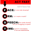
When a brain aneurysm ruptures, it causes bleeding into the subarachnoid space, otherwise known as a subarachnoid hemorrhage.

As the blood seeps into the skull, sudden symptoms can occur all at once that require immediate medical attention. These include:

  • Loss of consciousness
  • Vomiting/Nausea
  • Stiff Neck
  • Sensitivity to light
  • Dizziness or sudden trouble walking
  • Sudden numbness and weakness
  • Sudden severe headache
  • Sudden blurred or double vision
  • Drooping eyelid
  • Sudden pain above/behind the eye or difficulty seeing
  • Sudden change in mental status or awareness
  • Seizure

A ruptured brain aneurysm can cause a stroke, brain damage, or even death.

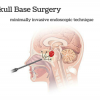
Approximately 15% of patients that suffer from subarachnoid hemorrhage die before reaching the hospital, while four out of seven who recover are faced with disabilities. Though the statistics are grim, Dr. Ramin Rak and the skilled surgeons at Neurological Surgery, P.C. are able to administer advanced surgical treatment to provide patients with the best care possible.


Related Article: http://raminrak.com/aneurysm-coiling/

Schwannoma

Thank you for visiting my blog. My name is Dr. Ramin Rak and I am a surgeon with Neurological Surgery, P.C.

I specialize in treating complex spinal and brain complications at the eleven different medical facilities affiliated with Neurological Surgery, P.C. and I am a member of the organization’s Neurosurgeon Brain Tumor Team.

The Brain Tumor team includes an Endovascular Neuroradiologist, two Neuro-oncologists, a Neuropsychologist, myself, and seven other neurosurgeons:

  • Dr. Michael H. Brisman, M.D., F.A.C.S.
  • Dr. Jeffrey A. Brown, M.D., F.A.C.S.
  • Dr. Lee Eric Tessler, M.D., F.A.A.N.S.
  • Dr. Alan Mechanic, M.D., F.A.C.S.
  • Dr. Robert N. Holtzman, M.D.
  • Dr. Vladimir Dadashev, M.D.
  • Dr. Gerald M. Zupruk, M.D., F.A.A.N.S.

The rest of the Brain Tumor Team and I work to treat brain tumors and complications caused by tumors, including gioblastoma, medulloblastoma, acoustic neuroma, and schwannoma.

Schwannoma is a homogenous tumor that is made up entirely of Schwann cells, which conduct nervous impulses along axons, provide trophic support for neurons, and support nerve development and regeneration.

The tumor cells are always on the exterior of the nerve though the tumor itself can cause nerve damage by pushing the nerve aside or into bone. Schwannomas become malignant in less than 1% of cases and are slow growing, but should still be treated if weakness numbness, pain or other symptoms are seen.

The Brain Tumor Team at NSPC uses surgery or stereotactic radiosurgery to treat schwannomas with radiosurgery being an option when the tumor is located in the head or spine.

The most common treatment is stereotactic radiosurgery, or the use of a machine called the Gamma Knife to focus high-powered x-rays at the spot of the tumor. Between 80 and 90% of the patients who opt instead to have surgery report that pain, weakness, and numbness disappears following the procedure.

Learn more about how the Brain Tumor Team treats schwannomas here.

Thank you for reading,

Dr. Ramin Rak


Ramin Rak Schwannoma

This is an example of a schwannoma. It typically has dense areas called Antoni A (black arrow) and looser areas called Antoni B (blue arrows). The cells are elongated (spindle shaped) and the nuclei have a tendency to line up as you see here in the Antoni A area. (via ucsf.edu)