Skull Base Surgery

Ramin Rak is a board-certified neurosurgeon with years of experience specializing in complex tumors, spine treatments, awake craniotomies, and skull base surgeries.

Skull base surgery is a specialized type of surgery performed to treat tumors and other diseases occurring in certain areas of the skull such as behind the eyes or in the nasal cavity. The skull is made up of bones and cartilage, forming the face and the cranium. The bones that form the base of the cranium also form the eye socket, some of the sinuses, the roof of the nasal cavity, as well as the bones surrounding the inner ear. The base of the skull is a very complex area where many vital parts of the body pass through including blood vessels, nerves, and the spinal cord.

Ramin Rak | Skull Base Surgery

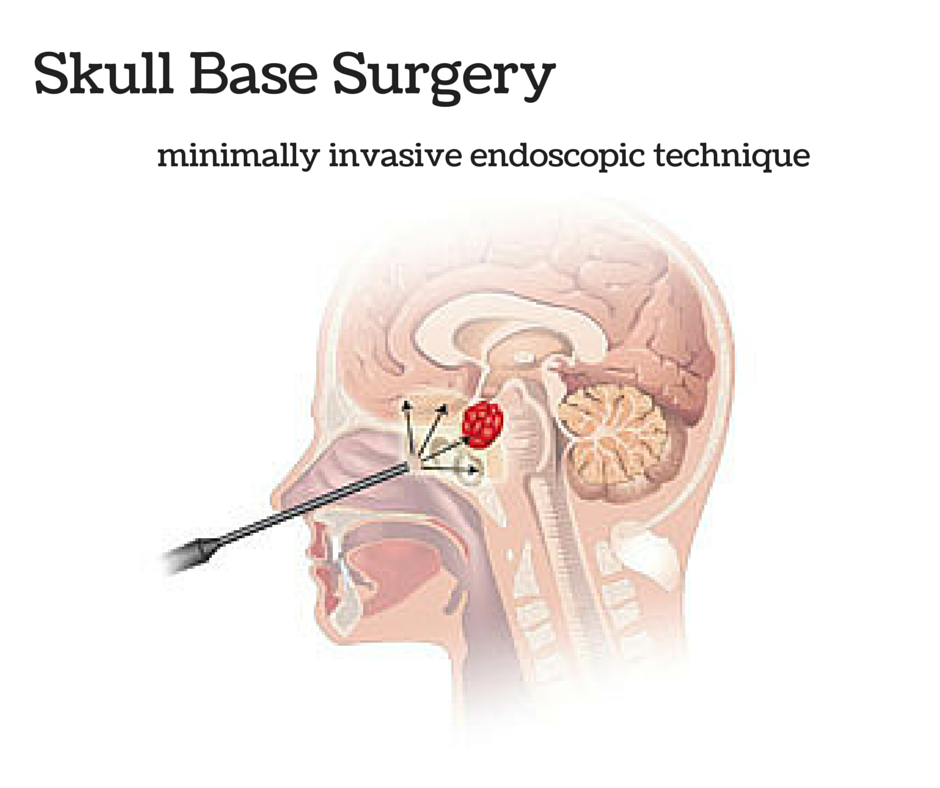
Ramin Rak uses the latest in minimally invasive techniques to perform skull base surgery.

Skull Base Surgery requires a multidisciplinary approach often involving ear, nose, and throat surgeons, neurosurgeons, plastic surgeons, radiologists, pathologists, oncologists, and other specialists. There are two main ways to perform skull base surgery. It may be done through a minimally invasive endoscopic procedure. This involves making a small incision through the natural openings of the skull, normally the nose or mouth. A small hole may also be made just above the eyebrow. Through this method, a neurosurgeon is able to remove the growth through a thin lighted tube referred to as an endoscope. An MRI picture of the skull base will usually be taken during this process to ensure that the entire growth has been removed successfully. The second way to perform skull base surgery is often referred to as traditional or open skull base surgery. This procedure requires making an opening in the skull. Parts of bone may be removed in order to reach the growth and have it properly removed.

Listed below are some of the common growths and conditions that may be treated with skull base surgery:

  • Growths caused by infections
  • Pituitary tumors
  • Sinus tumors
  • Glomus tumors
  • Cysts developed from birth
  • Cerebrospinal fluid leak
  • Meningiomas
  • Chordomas
  • Trigeminal neuralgia
  • Acoustic neuroma

Schwannoma

Thank you for visiting my blog. My name is Dr. Ramin Rak and I am a surgeon with Neurological Surgery, P.C.

I specialize in treating complex spinal and brain complications at the eleven different medical facilities affiliated with Neurological Surgery, P.C. and I am a member of the organization’s Neurosurgeon Brain Tumor Team.

The Brain Tumor team includes an Endovascular Neuroradiologist, two Neuro-oncologists, a Neuropsychologist, myself, and seven other neurosurgeons:

  • Dr. Michael H. Brisman, M.D., F.A.C.S.
  • Dr. Jeffrey A. Brown, M.D., F.A.C.S.
  • Dr. Lee Eric Tessler, M.D., F.A.A.N.S.
  • Dr. Alan Mechanic, M.D., F.A.C.S.
  • Dr. Robert N. Holtzman, M.D.
  • Dr. Vladimir Dadashev, M.D.
  • Dr. Gerald M. Zupruk, M.D., F.A.A.N.S.

The rest of the Brain Tumor Team and I work to treat brain tumors and complications caused by tumors, including gioblastoma, medulloblastoma, acoustic neuroma, and schwannoma.

Schwannoma is a homogenous tumor that is made up entirely of Schwann cells, which conduct nervous impulses along axons, provide trophic support for neurons, and support nerve development and regeneration.

The tumor cells are always on the exterior of the nerve though the tumor itself can cause nerve damage by pushing the nerve aside or into bone. Schwannomas become malignant in less than 1% of cases and are slow growing, but should still be treated if weakness numbness, pain or other symptoms are seen.

The Brain Tumor Team at NSPC uses surgery or stereotactic radiosurgery to treat schwannomas with radiosurgery being an option when the tumor is located in the head or spine.

The most common treatment is stereotactic radiosurgery, or the use of a machine called the Gamma Knife to focus high-powered x-rays at the spot of the tumor. Between 80 and 90% of the patients who opt instead to have surgery report that pain, weakness, and numbness disappears following the procedure.

Learn more about how the Brain Tumor Team treats schwannomas here.

Thank you for reading,

Dr. Ramin Rak


Ramin Rak Schwannoma

This is an example of a schwannoma. It typically has dense areas called Antoni A (black arrow) and looser areas called Antoni B (blue arrows). The cells are elongated (spindle shaped) and the nuclei have a tendency to line up as you see here in the Antoni A area. (via ucsf.edu)

Gamma Knife Procedure

Hello readers, Ramin Rak here with yet another blog post about a complex neurosurgical method that I have used to treat patients.

Gamma Knife is a procedure, like the CyberKnife System, that allows doctors like me to treat tumors and ailments without having to resort to surgery. Gamma Knife surgery was approved for use by the US Food and Drug Administration (FDA) thirty years ago and I am one of the few New York area neurosurgeons certified in this technique.

When I use Gamma Knife procedures for a patient, I can deliver over 200 beams of radiation right to the tumors and lesions.

Unlike with traditional radiation, patients do not experience the regular side effects because such low doses of radiation are injected. Patients can be given several shots of radiation during one session, and sessions can be repeated until the tumor has stopped growing.Gamma knife procedures can be used to treat:

  • Benign tumors, such as meningiomas, schwannomas, craniopharyngiomas, pineal tumors, acoustic neuromas, and pituitary adenomas.
  • Malignant tumors, such as metastatic tumors, chordomas, ependymomas, medulloblastomas, astrocytomas, and anaplastic astrocytomas.
  • Arteriovenous malformations (ATV)
  • Trigeminal neuralgia
  • Parkinson’s Disease

Generally Gamma Knife use is restricted to those tumors that are less than 4 centimeters in size.

This procedure has a success rate between 90 and 95 percent, as that is how often the procedure stops tumor growth. The majority of the time it also causes tumors to shrink, taking anywhere from one week to a year to stop growth.

When a patient undergoes Gamma Knife treatment, he or she will be fitted with a clear, plastic frame.  My team uses 3D computer imaging to determine where radiation must be aimed to successfully reach the tumor. Patients are then fitted with the Gamma Knife apparatus, which is a metal helmet that emits radiation beams. The apparatus then directs radiation beams to the tumor.

This is how I complete Gamma Knife treatments. If you have any questions, check to see if I have answered them on Quora, or reach out to me directly.

Thanks for reading,

Ramin Rak