Microdiscectomy

Hi all, Ramin Rak here with another blog post about the complex neurosurgical procedures I complete at Neurological Surgery, P.C.

I specialize in the completion of surgeries meant to treat ailments affecting the brain and spine. In previous blog posts I have discussed my work with the NSPC Brain Tumor Center and spinal procedures such as Spinal Fusion and the X-Stop Procedure. One spinal procedure that I have not written about is a microdiscectomy.

When a patient complains of leg pain I can use magnetic resource imaging (MRI) or a computer tomography (CT) scan to determine if the patient has a herniated disc.

Using these results, I then decide whether or not a microdiscectomy will relieve the patient’s symptoms.

This spinal procedure is primarily used to treat leg pain, specifically leg pain arising from a herniated lumbar disc. Compression or impingement on the nerve root, defined as bone colliding with or striking the nerve root, will cause considerable leg pain. A microdiscectomy is used to relieve leg pain, and in many cases the patient will feel relief immediately after completion of surgery.

When completing a microdiscectomy, I examine the disc and nerves under a high powered microscope so that I only need to make a small incision.

By using this microneurosurgical technique instead of performing a discectomy, patients experience a much smaller recovery time because there is less tissue damage.

After the incision is made, I move the back muscles off of the bony arch (lamina) of the spine. I am then able to enter the spine by removing a membrane over the nerve roots and visualize the nerve using operating glasses. Once herniated disc material is removed, the procedure is complete.

For more information about the types of spinal procedures I have completed, take a look at my other blog posts or view some of the surgical videos I have uploaded to Vimeo by clicking here.

Thanks for reading,

Ramin Rak


PS For more information on the completion of a microdiscectomy, visit this link.

Kyphoplasty Surgery

Hello again, my name is Ramin Rak and I am writing this post to share information about another complex spinal procedure that I perform at Neurological Surgery, P.C.: Kyphoplasty Surgery.

I am one of six Neurological Surgery, P.C. neurosurgeons who performs kyphoplasty surgery. The others are:

  • Dr. Stephen D. Burstein
  • Dr. William J. Sonstein
  • Dr. Benjamin R. Cohen
  • Dr. Artem Y. Vaynman
  • Donald S. Krieff, D.O., F.A.C.O.S.

I perform kyphoplasty surgery in order to reverse spinal compression caused by a spinal bone fracture.

Individuals who experience compression lose vertebral body height and experience intractable pain. Fortunately kyphoplasty surgery removes pain relief roughly 48 hours after completion of the procedure, and patients can leave the hospital on the same day the procedure is completed.

Once the patient has been sedated, I make a small incision in the patient’s back so that I can insert a narrow tube-like needle into the fractured vertebral body.

I then use an imaging technique called fluoroscopy, which uses x-rays to provide a real-time moving image of the patient’s spinal structure, to guide the needle into the fractured area. Once a path has been made to the spot of the fracture, I insert a balloon into the tube, guide it to the vertebrae, and then slowly inflate it. The inflated balloon elevates the spinal structure, which restores vertebral body height. Next I remove the balloon and fill the cavity created by the balloon with a cement-like material that hardens quickly and stabilizes the spinal structure.

The entire procedure takes roughly one hour per affected vertebra and following conclusion of the procedure, the patient is observed in the recovery room until my doctors determine that he or she can leave.

This is how I complete kyphoplasty surgery at Neurological Surgery, P.C.

Learn more about how I complete this procedure by reaching out to me on Doctor’s Hangout.

Thanks for reading,

Ramin Rak


Ramin-Rak-Kyphoplasty

During kyphoplasty surgery, a balloon is inserted into the spine and inflated via a small tube.

Schwannoma

Thank you for visiting my blog. My name is Dr. Ramin Rak and I am a surgeon with Neurological Surgery, P.C.

I specialize in treating complex spinal and brain complications at the eleven different medical facilities affiliated with Neurological Surgery, P.C. and I am a member of the organization’s Neurosurgeon Brain Tumor Team.

The Brain Tumor team includes an Endovascular Neuroradiologist, two Neuro-oncologists, a Neuropsychologist, myself, and seven other neurosurgeons:

  • Dr. Michael H. Brisman, M.D., F.A.C.S.
  • Dr. Jeffrey A. Brown, M.D., F.A.C.S.
  • Dr. Lee Eric Tessler, M.D., F.A.A.N.S.
  • Dr. Alan Mechanic, M.D., F.A.C.S.
  • Dr. Robert N. Holtzman, M.D.
  • Dr. Vladimir Dadashev, M.D.
  • Dr. Gerald M. Zupruk, M.D., F.A.A.N.S.

The rest of the Brain Tumor Team and I work to treat brain tumors and complications caused by tumors, including gioblastoma, medulloblastoma, acoustic neuroma, and schwannoma.

Schwannoma is a homogenous tumor that is made up entirely of Schwann cells, which conduct nervous impulses along axons, provide trophic support for neurons, and support nerve development and regeneration.

The tumor cells are always on the exterior of the nerve though the tumor itself can cause nerve damage by pushing the nerve aside or into bone. Schwannomas become malignant in less than 1% of cases and are slow growing, but should still be treated if weakness numbness, pain or other symptoms are seen.

The Brain Tumor Team at NSPC uses surgery or stereotactic radiosurgery to treat schwannomas with radiosurgery being an option when the tumor is located in the head or spine.

The most common treatment is stereotactic radiosurgery, or the use of a machine called the Gamma Knife to focus high-powered x-rays at the spot of the tumor. Between 80 and 90% of the patients who opt instead to have surgery report that pain, weakness, and numbness disappears following the procedure.

Learn more about how the Brain Tumor Team treats schwannomas here.

Thank you for reading,

Dr. Ramin Rak


Ramin Rak Schwannoma

This is an example of a schwannoma. It typically has dense areas called Antoni A (black arrow) and looser areas called Antoni B (blue arrows). The cells are elongated (spindle shaped) and the nuclei have a tendency to line up as you see here in the Antoni A area. (via ucsf.edu)

NSPC Brain Tumor Center

NSPC Brain Tumor Center with Ramin Rak

Hi all, Ramin Rak here with another blog post about my work as a neurosurgeon.

I am affiliated with Neurological Surgery, P.C., New York’s private largest neurosurgical group. One of the resources that Neurological Surgery, P.C. uses to provide better treatment is the Long Island Brain Tumor Center (their official website is located here).

I am one of the neurosurgeons who is part of the Neurosurgeon Brain Tumor Team, along with:

  • Dr. Michael H. Brisman, M.D., F.A.C.S.
  • Dr. Jeffrey A. Brown, M.D., F.A.C.S.
  • Dr. Lee Eric Tessler, M.D., F.A.A.N.S.
  • Dr. Alan Mechanic, M.D., F.A.C.S.
  • Dr. Robert N. Holtzman, M.D.
  • Dr. Vladimir Dadashev, M.D.
  • Dr. Gerald M. Zupruk, M.D., F.A.A.N.S.

The Brain Tumor Team also includes an Endovascular Neuroradiologist (Dr. John Pile-Spellman, M.D.), a Neuropsychologist (Dr. Gad E. Klein, Ph. D.), and two neuro-oncologists (Dr. Paul Duic, M.D. and Dr. Jai Grewal, M.D.).

The Long Island Brain Tumor Center focuses on treating conditions like acoustic neuroma, schwannoma, ependymoma, astrocytoma, and other ailments related to brain tumors.

A brain tumor occurs as a result of uncontrolled cell division in the brain itself, glial cells, lymphatic tissue, the cranial nerves, brain envelopes, the skull, pituitary and pineal gland, or can even be spread from cancer located in other areas. Symptoms of a brain tumor include phantom odors and tastes, while onset symptoms like an epileptic seizure in a patient with no history of epilepsy or sudden intracranial hypertension have also been observed.

The team of neurosurgeons within the Long Island Brain Tumor Center diagnoses tumors using computed tomography (CT) and magnetic resonance imaging (MRI) mapping techniques.

Tumors must be investigated before a diagnosis and treatment options can be determined, but the good news is that not all brain tumors are cancerous.

Take a look at my other blog posts to find out how I use techniques like Gamma Knife, and CyberKnife procedures to treat brain tumors without surgery.

Thanks for reading,

Ramin Rak

Spinal Fusion

Good afternoon, and thank you for visiting my blog.

My name is Dr. Ramin Rak and I am a neurosurgeon with Neurological Surgery, P.C. Though I primarily specialize in techniques that treat brain injuries and tumors (like the Cyber Knife and GammaKnife procedures that I spoke of in earlier blog posts), I also complete complex procedures intended to alleviate spinal conditions. Last week I detailed my use of the X-Stop Procedure for individuals suffering from back pain and this week I will talk about my technique for treating a condition called spondylolisthesis.

Spondylolisthesis occurs when a damaged or fractured joint allows a vertebra to slip forward and pinch a nerve joint in the spine.

This pain will travel all the way down to the patient’s legs via the sciatic nerve. Surgeons perform a spinal fusion to treat this condition and alleviate patient pain. At Neurological Surgery, P.C. I am a member of the Spinal Fusion Team along with:

  • William J. Sonstein, M.D. F.A.C.S.
  • Benjamin R. Cohen, M.D. F.A.C.S.
  • Artem Y. Vaynman, M.D. F.A.A.N.S.
  • Donald S. Krieff, D.O. F.A.C.O.S.

My team and I perform the spinal fusion procedure to treat intractable back pain by fusing two or three lumbar spine segments.

We start by removing the lamina, which is the portion of the spine that covers the problematic vertebra. Next, the Spinal Fusion team removes any piece of bone that is pinching the vertebra, relieving any pressure and pain. A bone graft is then performed, which fuses together the two spine segments around the vertebra. Finally, screws and rods are used to hold the spine in place while the grafts heal. Once the grafts heal, they will fuse into place and keep the discs from slipping and causing future complications.

Learn more about the spinal fusion team and the PLIF (posterior lumbar interbody fusion) or spinal fusion procedure by clicking here to view a video demonstration on NSPC’s website. Stay up to date on my other neurosurgical techniques when you follow me on Twitter.

Thanks for reading,

Dr. Ramin Rak


Ramin Rak Spinal Fusion

Screws and rods are used to hold the bone grafts in place so that the spinal fusion results are permanent.

The X-Stop Procedure

Hello all, Dr. Ramin Rak here with another blog post.

Many of my posts have focused on complex neurosurgical techniques that I have used and still use, but I also specialize in completing complex spinal surgeries for individuals suffering from back pain. One of my specialties is the X-Stop Procedure, and I am one of only seven surgeons at Neurological Surgery, P.C. who complete this procedure. The others are Dr. Stephen D. Burnstein, Dr. William Sonstein, Dr. Benjamin Cohen, Dr. Artem Vaynman, Dr. Alan Mechanic, and Dr. Donald Krieff.

The X-Stop Procedure involves the insertion of a titanium implant called the X-Stop Spacer.

The spacer is placed in order to treat back pain and leg pain caused by lumbar spinal stenosis (LSS). LSS takes place when space between the vertebrae is reduced, as this lack of space causes bone or tissue to come in contact with the spinal nerve. Individuals who are suffering from LSS find relief when seated or bending over because these positions open up space between the vertebrae. The X-Stop Procedure draws inspiration from this concept, using the X-Stop Spacer to lift the vertebrae off of the pinched nerve and provide long-term relief.

X-Stop Spacer

A diagram of the X-Stop Spacer following placement.

I complete the minimally invasive X-Stop Procedure by surgically inserting the X-Stop Spacer between two bones in the back of the spine; usually L3/4 or L4/5.

The surgical site is located between the posterior spinous processes, which is an area you can feel for yourself by running a finger along your spine. The X-Stop Spacer stays in place by working with one’s natural spinal anatomy, which means that the procedure is completed without using screws or needing to be attached to bones or ligaments.

The X-Stop Procedure takes between 50 and 90 minutes, and patients are able to walk around shortly after the anesthesia wears off. Patients see results immediately after the procedure, as they will no longer experience back pain when standing or walking around.

Learn more about the X-Stop Procedure by visiting their website, or leave a comment below if you still have questions.

Thank you for reading,

Dr. Ramin Rak

My Second Awake Craniotomy

Good afternoon and welcome back to my blog.

My name is Ramin Rak, I am a neurosurgeon with Neurological Surgery, P.C. in New York and one of my specialties is performing awake craniotomies.

In July of 2010, I received media attention after completing the first awake craniotomy that had ever been performed at the North Shore-LIJ Huntington Hospital.

The procedure was notable for me because it was only my second awake craniotomy.

The need for the procedure was uncovered after 23-year-old Boris Arrazia experienced a seizure while driving his car. Arrazia’s passenger was able to navigate the car to the side of the road and put it in park, and a passerby performed CPR and then called for help.

Arrazia was brought to Huntington Hospital where doctors uncovered the cause of the stroke: a tumor located in the temporal lobe of his brain.

Due to the tumor’s unusual location, the tumor did not show up when using advanced imaging techniques. Because the tumor was right on his speech area, there was no way to map and understand how we are going to control the reception of the brain without the patient being awake. By performing an awake craniotomy, my team was able to make sure we did not damage areas of the brain dealing with speech during surgery by asking Arrazia to repeat phrases or describe pictures. Arrazia recounts:

“I was nervous when Ramin Rak first told me the diagnosis, but happy to hear that the tumor was operable,” Arriaza said. “Ramin Rak told me that I would be conscious for the surgery, and that it might be uncomfortable and possibly a little painful. During the surgery, they showed me pictures and asked me to name the objects [like] a horse [or] a table.”

Though there are a lot of risks associated with awake craniotomies, including serious bleeding, there were no complications seen while removing Arrazia’s tumor. Four days later, he walked out of the hospital and reunited with his family.

Learn more about this particular awake craniotomy by taking a look at the media coverage this procedure received:

http://www.northshorelij.com/NSLIJ/Islip+Man+Undergoes+Brain+Surgery+While+Awake  

http://huntington.patch.com/articles/huntington-hospital-doc-performs-awake-craniotomy


If you are interested in learning more about how I perform awake craniotomies, visit my Quora profile.

Thank you for reading,

Ramin Rak

Ramin Rak awake craniotomy

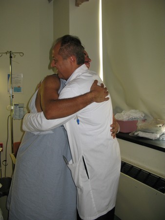
Boris Arrazia thanks Ramin Rak following completion of the awake craniotomy.