Symptoms of a Stroke

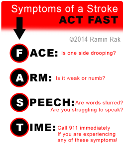
F.A.S.T. Stroke Symptoms

Ramin Rak breaks down the F.A.S.T. stroke symptoms.

Ramin Rak practices his advanced knowledge of microneurosurgical techniques at Neurological Surgery P.C. on Long Island.

Here, he specializes in awake craniotomies, spinal surgeries, skull-based surgeries, and treatment of tumors.

More information on these complex procedures can be accessed through his Pinterest page.

A stroke is caused by a disturbance in the blood supply and results in the loss of function.

An ischemic stroke is caused by a clot that obstructs blood flow, while a hemorrhagic stroke is a result of a rupturing blood vessel. A transient ischemic attack can also occur, which is essentially a mini stroke caused by a temporary clot. Both types of strokes are extremely serious, as they are the number four cause of death, and the leading cause of adult disability in the United States.

According to strokeassociation.org, F.A.S.T is an easy way to remember the sudden signs of a stroke:

  • Face Drooping- If one side of your face is drooping or your smile is uneven, it could indicate a stroke.
  • Arm Weakness- When one arm is weak, numb, or drifts downward when your arms are raised, seek immediate medical attention.
  • Speech Difficulty- If someone is struggling to form words or unable to repeat a simple sentence, a stroke may be the cause.
  • Time to call 911- If you are experiencing any of these symptoms, call 911 immediately, even if the symptoms go away.

Beyond these core symptoms, a person may also present numbness of the leg, arm or face; trouble seeing; an unexplained sudden, severe headache; dizziness or loss of coordination; and confusion or trouble understanding.

Quick stroke treatment can save lives, so it is crucial to seek immediate medical attention if you have shown any of the above symptoms. After a doctor makes a diagnosis, they will begin a treatment plan specific to the cause of the stroke.
Good Bedside Manner

Bedside Manors Ramin Rak is a leading Neurosurgeon with a long history of expert specialties and training in micro-neurosurgical procedures. He has performed countless surgeries, and specialized in highly-complex awake craniotomies, skull-based surgeries, spine treatments and tumors. Ramin Rak attended the Free University of Brussels for his medical degree, and previous to this, for his undergraduate degree with high honors in Medical Basic Sciences. Ramin Rak has, since this time, gained a considerable amount of training, expertise, which has helped him gain effective bedside manner. While the actual procedural and administrative aspects of a successful surgery are vital to a smooth surgery for a patient, knowing what a good bedside manner is and how to employ it, is arguably just as important. Here are some tips for gaining and employing a good bedside manner to quell fears in your patients and make the entire process as comfortable and positive as possible.

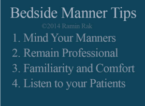
  1.       Mind Your Manners: First rule of thumb in presenting the best bedside manner-which should be included in any profession- is using good manners at all times, with the necessary please and thank yous, as well as an overall gesture of respect.
  2.       Professionalism: As a doctor, you should always be well-groomed, confident, assured, and knowledgeable.
  3.       Familiarity: Make sure that you patients feel like you care about them specifically, You may deal with 100 patients a day, but you should address each singularly with names, histories, and the care that each patient deserves. Doing so will bring more comfort to each patient more than you might think.
  4.       Listening: Yes-you are the more knowledgeable about medicine in the room, probably, but listening to what your patients are feeling, their fears, symptoms, and questions is a vital part both to better understanding how to best treat them, but also, how to make them feel like an important part of the process (and they are).

Learn more about Ramin Rak on his About.me page.

Glioblastoma

GBM

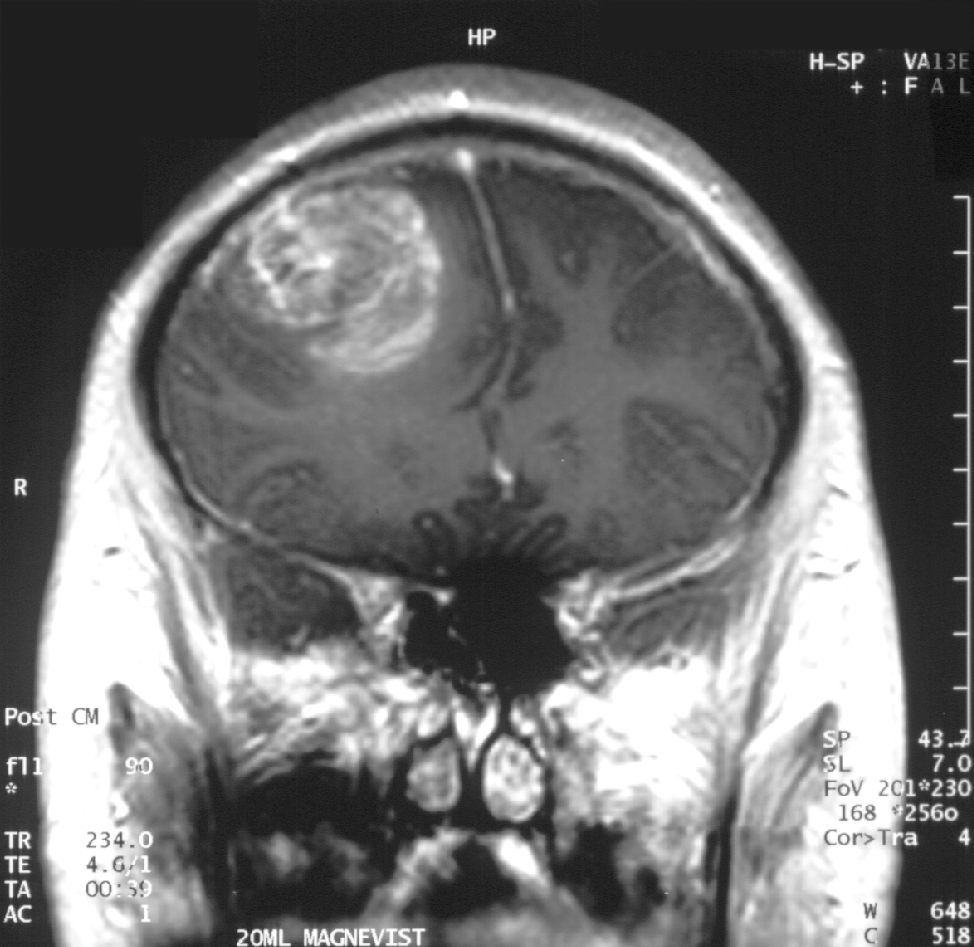
Glioblastoma in an x-ray. (Source: Wikipedia.org)

Hello again, this is Ramin Rak, back with another post about a medical procedure I performed at Neurological Surgery, P.C. in Long Island.

I specialize in treating disorders related to the brain and spine, and recently removed a glioblastoma multiforme tumor during a high-profile surgery.

Glioblastomas (GBM) are tumors found in the cerebral hemisphere or spinal cord.

These tumors are highly cancerous due to their ability to reproduce cells quickly. In addition, they are supported by a large network of blood vessels. The cause of these tumors are unknown but due to their rapid growth, symptoms commonly involve pressure on the brain. Headache, vomiting, nausea, and drowsiness are the most common.  Treating GBM is very difficult because these tumors contain many different types of cells. Furthermore, the network of tentacles in the brain make it extremely difficult to remove these tumors without affecting the patient’s language and coordination. For these reasons, your surgeon may decide to combine several approaches to treat your GBM.

Recently, Donald Squire, 52, of East Northport was diagnosed with a GBM that required surgery.

His only symptom was a twitching in one of his eyes. His wife suspected a stroke but brain scans revealed that the twitching was due to a tumor. Fortunately, I was able to use proprietary technology to enter the inner labyrinths of his brain and remove the tumor. To reach the tumor, I used brain-mapping technology and neuronavigation, which made it possible to move eloquently throughout the delicate areas of his brain. The patient was awake during surgery in order for me to monitor his language, vision, and speech. After the surgery, I had concerns that Squire might lose some of his peripheral vision, but he hasn’t noticed any changes. Squire is very fortunate to have had access to this technology, as not too long ago, this area of his brain would have been unreachable using conventional techniques.

You can learn more about Squire’s procedure here: http://grow-your-practice.net/wp-content/uploads/2013/07/Dr-Rak-Newsday-062213.pdf


Thanks for reading,

Ramin Rak

You can learn more about me and my specialties in the Commack Patch, or by connecting with me on Doctor’s Hangout: www.doctorshangout.com/profile/DrRaminRakMD

Gamma Knife Procedure

Hello readers, Ramin Rak here with yet another blog post about a complex neurosurgical method that I have used to treat patients.

Gamma Knife is a procedure, like the CyberKnife System, that allows doctors like me to treat tumors and ailments without having to resort to surgery. Gamma Knife surgery was approved for use by the US Food and Drug Administration (FDA) thirty years ago and I am one of the few New York area neurosurgeons certified in this technique.

When I use Gamma Knife procedures for a patient, I can deliver over 200 beams of radiation right to the tumors and lesions.

Unlike with traditional radiation, patients do not experience the regular side effects because such low doses of radiation are injected. Patients can be given several shots of radiation during one session, and sessions can be repeated until the tumor has stopped growing.Gamma knife procedures can be used to treat:

  • Benign tumors, such as meningiomas, schwannomas, craniopharyngiomas, pineal tumors, acoustic neuromas, and pituitary adenomas.
  • Malignant tumors, such as metastatic tumors, chordomas, ependymomas, medulloblastomas, astrocytomas, and anaplastic astrocytomas.
  • Arteriovenous malformations (ATV)
  • Trigeminal neuralgia
  • Parkinson’s Disease

Generally Gamma Knife use is restricted to those tumors that are less than 4 centimeters in size.

This procedure has a success rate between 90 and 95 percent, as that is how often the procedure stops tumor growth. The majority of the time it also causes tumors to shrink, taking anywhere from one week to a year to stop growth.

When a patient undergoes Gamma Knife treatment, he or she will be fitted with a clear, plastic frame.  My team uses 3D computer imaging to determine where radiation must be aimed to successfully reach the tumor. Patients are then fitted with the Gamma Knife apparatus, which is a metal helmet that emits radiation beams. The apparatus then directs radiation beams to the tumor.

This is how I complete Gamma Knife treatments. If you have any questions, check to see if I have answered them on Quora, or reach out to me directly.

Thanks for reading,

Ramin Rak


Neuronavigation

Hi all, Ramin Rak here again with another blog post.

I am a neurological surgeon affiliated with Neurological Surgery, P.C. in New York. I specialize in using microneurosurgical techniques for the treatment of brain tumors and complex spine diseases. I also perform skull-based surgery and awake craniotomies.

Recently at Huntington Hospital, which is part of the North Shore-Long Island Jewish Hospital System, I had a chance to combine neuronavigation and microsurgery to treat a patient named Roger Sencer.

While out driving, Roger suddenly forgot where he was, who he was, and even forgot recent events in his life. He was brought to Huntington Hospital where he was diagnosed with a large tumor resting on the brain: a condition called meningioma.

The next day I met with Roger and we bonded quickly over our interest in spiritual matters.

The surgery took nearly 12 hours but luckily at the end of surgery I was confident that he had been cured. Roger’s wife explains the wait below in this excerpt from an article from Huntington Hospital’s website:

Jane, on the other hand, remembers every detail – the nearly 12 hours in the surgical waiting room surrounded by friends and loved ones, being impressed by the technology in place throughout the hospital, the compassion of the entire hospital staff, and most importantly, the serene look on Dr. Ramin Rak’s face when he finally emerged from the operating room and said, “This is one of those times when I can confidently say the patient is cured.” Their entire contingency of family and friends erupted in applause.

The surgery took so long because Roger’s tumor was at the base of the skull surrounded by essential neurovascular structures.

Using pre-operative navigation planning the day before the surgery, I was able to create a three-dimensional map of his head. This allowed me to avoid injury to blood vessels and preserve the olfactory nerve during surgery.

Fortunately, neuronavigation was possible thanks to this map and my neurosurgical techniques.

Three months later Roger returned to work.

In the future I will speak more about some of the intricate surgeries I have performed.

Thank you for reading,

Ramin Rak

Ramin Rak Neuronavigation