Aneurysm Coiling

Hi all, Ramin Rak here with another blog post about a complicated neurosurgical procedure I perform at Neurological Surgery, P.C.: aneurysm coiling.

I primarily treat conditions in the brain and spine so I only perform aneurysm coiling to address brain aneurysms. Endovascular aneurysm coiling is one of two techniques (along with open surgical clipping) that can be used to treat brain aneurysms, but sometimes a physician will choose to closely observe an aneurysm instead of recommending one of these two treatments.

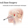
Aneurysm coiling is a minimally invasive endovascular procedure performed to treat an aneurysm, which is a balloon-like bulge of an artery wall (learn more).

As an aneurysm grows, it will thin and weaken until it becomes so thin that it leaks or ruptures. A ruptured aneurysm will release blood into the space around the brain, called a subarachnoid hemorrhage, and is life threatening. Treatment involves stopping blood from flowing into the aneurysm but still allowing blood to flow freely through the normal arteries. While open surgical clipping accomplishes this from the outside, aneurysm coiling does so from the inside.

Ramin Rak Aneurysm Coiling

This diagram depicts the insertion of small platinum coils into the aneurysm using a catheter

I begin the procedure by giving the patient anesthesia while they are on the x-ray table.

Next, I locate the femoral artery and use a needle to insert a long plastic tube (the catheter) into the bloodstream. Dye is then injected through the catheter to make blood vessels visible on my x-ray monitor, allowing me to guide the catheter to one of four arteries in the neck that lead to the brain. After the catheter is placed, I take x-ray photos and use them to take measurements of the aneurysm.

A second smaller catheter travels through the first catheter and makes its way to the aneurysm itself.

Small platinum coils are then passed through the catheter until they emerge in the aneurysm, and this process continues until the aneurysm is completely packed with coils. I then inject contrast agent so that I can confirm that blood is no longer flowing into the aneurysm and finally close the puncture site in the artery.

Endovascular aneurysm coiling has a long-term success rate between 80 and 85%.

Thanks for reading,

Ramin Rak

Learn more about my specialized neurosurgical procedures on Brand Yourself: http://raminrak.brandyourself.com/ or visit my WordPress blog.

Advanced Awake Craniotomy Technologies

Hi all, Ramin Rak here with the latest news about a medical procedure I perform called awake craniotomy.

At Neurological Surgery, P.C. in Long Island, New York, I am one of the leading neurosurgeons performing this procedure. When I conduct an awake craniotomy, I work alongside a team of skilled physicians to operate some of today’s  most advanced neuro-navigational technology. My expertise in micro-neurosurgical techniques has allowed me to successfully establish awake craniotomy programs in multiple hospitals around the Long Island area.

Ramin Rak awake craniotomy

“My colleague tests a patient’s motor skills during an awake craniotomy.”

Awake craniotomy is a procedure in which patients remain awake during brain tumor surgery in order to determine whether the operation will affect specific areas of the brain that control speech, vision, and movement.

Once I identify a patient as a good candidate for an awake craniotomy, I work with the specialized team at Neurological Surgery, P.C. to establish a baseline of cognitive function and precisely pinpoint the tumor’s location. The specialized team is composed of neurophysiologists (who monitor the brain’s electrical impulses), nurses, and physicians assistants to ensure everything runs smoothly. Before operating on a patient, I use a unique mapping approach to map the brain’s gray matter and nerve fibers within the white matter. Mapping techniques have helped determine where the most brain damage potential lies, allowing me to protect the patient’s ability to speak, move, and see.

Thanks to functional brain imaging (functional MRI) and other neurological technologies, I can perform awake craniotomies on patients whose tumors were previously thought to be inoperable.

A functional MRI scan is especially important because it identifies functional areas of the brain that are impacted by the tumors. Over the past couple of years, neuronavigation and new types of anesthesia have also made the procedure safer and easier to conduct. The most reassuring aspect that relieves my patients is when I tell them that they will not feel pain when they are either awake or sedated during an awake craniotomy.

For further information about awake craniotomy, read my full press release here: http://www.prweb.com/releases/2013/2/prweb10445390.htm or learn about other procedures I perform by reading my blog.


Thanks for reading,

Ramin Rak